Table of Contents
The Fundamental Unit of Life Activity Solution: Class 9 Science Chapter 5
Content:
The Fundamental Unit of Life Activity Solution
Activity 5.1
Activity 5.1 asks us to peel an onion and observe it under a simple microscope.
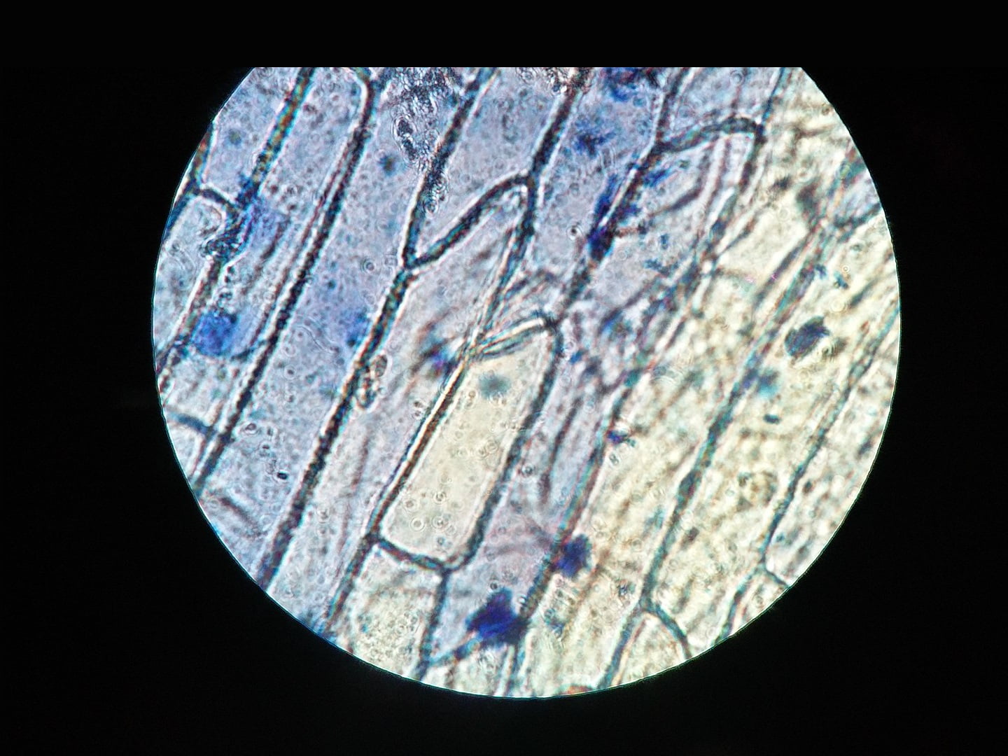
Some leaves and fruits like an onion peel, leaf skin etc have well-defined cell walls and nucleus. These cells are easily observable by the simple microscope. So, we use such cell to observe cell structures.
When we put such cells on microscope we see we defined cell wall and nucleus as given in the figure.
Class 9 Science Chapter 5 activity solution
Activity 5.2
Activity 5.2 asks us to see cells of different plants and part under the microscope.

Different cells have different shape and size, structure. These characteristics depend on the species e.g. Human cells different from that other organism; type of cells e.g. stem cells differ from leaf cells etc and functions.
Activity 5.3
Activity 5.3 asks us to peel off the outer shell of an egg using dilute hydrochloric acid and observe the size of the egg in pure water and distilled water.
Observation: Peeled egg in water swells and become bigger, while in saline water it shrinks in size.
Explanation:
The eggshell is made up of calcium carbonate. It reacts with dilute hydrochloric acid and form soluble compound (calcium chloride) which washes away. A cell membrane lies beneath the shell. A cell membrane is permeable to a few things while impermeable to others. When we put peeled egg into the water, it absorbs water and swells.
Saline water has more osmotic pressure than the inside of the egg. As a result, water from the inside of the egg rushes outwards and we see shrinkage in the size of the egg.
Class 9 Science Chapter 5 activity solution
Activity 5.4
Activity 5.4 is similar to activity 5.3. It asks us to do the same but with different materials like raisins and dried apricots.
Observation and explanation: Observation and explanation are similar to the shelled-off egg in water. In water raisins and dried apricot absorb water and swell in size.
Activity 5.5

Activity 5.5 asks us to know about an electron microscope.
Answer: From a simple microscope we can see only cell wall and nucleus. To see other organelles, cell wall and cell membrane in detail we need a more powerful microscope. For this, we use an electron microscope. In an electron microscope, a concentrated beam of electrons are thrown on to the cell and their reflection is recorded on the screen. This gives a very magnified image of the object. With this, we can easily see the composition of a cell wall, cell membrane, organelles etc.
The Fundamental Unit of Life Activity Solution
Activity 5.6
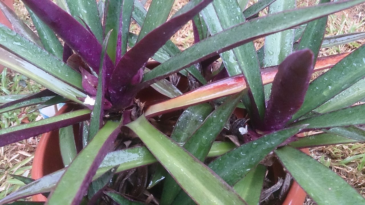
Activity 5.6 asks us to observe Rhoeo leaf under the microscope; compare the osmosis with its leaf boiled in water.
Observation and explanation:
Rhoeo is a common decorative plant found in our home and gardens. Its slides are easy to prepare, so we use them to study cell components and functions.
Under the microscope, we see changes similar to eggs, raisins and dried apricots i.e. swelling in water and shrinkage with saline or sugar solution.
Boiling of leaf breaks the cell wall and other cellular components like cytoplasm. Osmosis in a leaf is a function of protoplasm. As a result, we do not see any change in shape or size in leaf cells whether in water or solution.
Inference/conclusion:
A cell is alive until its protoplasm is functioning. A living cell responds to changes in external osmotic pressure. While a dead cell ceases to show response to osmotic pressure.
Activity 5.7

Observation and explanation:
A thin layer of mucosal cells lines the inside of the cheek cells. These are easy to scrape off and observe under the microscope.
Under high power microscope, we can see cell membranes, outlines of various organelles like Golgi bodies, endoplasmic reticulum, nucleus etc.
A nucleus is the most important part of the cell. It coordinates functions of the cells by protein synthesis. Under normal condition, it appears as a double-membraned structure with thick dense material inside, chromatin. When the cell is under the division process, the nucleus appears bigger with different chromosome visible.
Next:
- The Fundamental Unit of Life In-text and Exercise question Solution.
- The Fundamental Unit of Life MCQ – Objective Questions.
see also:
Ref: NCERT chapter 5.


Hi I love your vebsiste
Hsbsbbsbsnbns bye
Thank you
It helped a lot ✨
Very easy to understand thank you so much .
Very Useful sir.
Will you please explain the activities in detail for 9th bio as it is done for 10th bio .